Oogheelkundig Onderzoek


OCT, FAG, FAF, MAIA
Voor het vaststellen van maculadegeneratie test de oogarts eerst uw gezichtsscherpte.
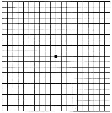
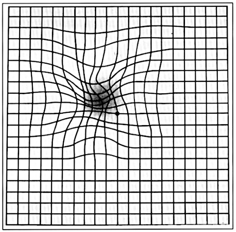
Verder kan men met een bladzijde met ruitjespatroon testen of er vervormingen of andere afwijkingen in het gezichtsvermogen optreden.
Dit wordt de Amslertest genoemd. Deze test is zeer geschikt voor zelfcontrole thuis.Indien u vervormingen waarneemt, dient u binnen een week door een oogarts te worden gezien.
Na verwijden van de pupil door het indruppelen van de ogen kan de oogarts met een lamp en een vergrootglas het volledige netvlies en in het bijzonder de macula onderzoeken. Dit onderzoek wordt "oogspiegelen" genoemd.
Wanneer de uitgebreidheid onvoldoende duidelijk is, is aanvullend onderzoek noodzakelijk, zoals Fluorescentie Angiografie (FAG), Autofluorescentie Onderzoek (FAF) en Optical Coherance Tomography (OCT).
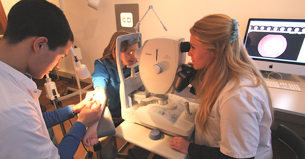
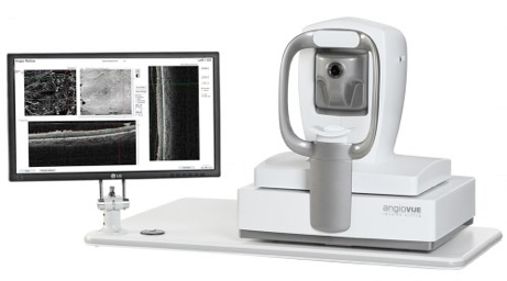
Als enige kliniek in Nederland zijn al onze spreekkamers standaard voorzien van een Optovue iVue of Zeiss Cirrus HD-OCT ( Optical Coherence Tomography ).
Hierdoor kan dit uiterst geavanceerde en noodzakelijke netvliesonderzoek bij maculadegeneratie direct tijdens het consult worden gedaan.
In alle andere klinieken wordt dit onderzoek op afspraak door een TOA (Technisch Oogheelkundig Assistent) verricht. Het kost hierdoor extra tijd om tot een diagnose te komen en het onderzoek heeft hierbij een lagere specificiteit en nauwkeurigheid dan wanneer het OCT onderzoek door uw oogarts zelf wordt gedaan.
Gezichtsscherpte en Amslertest
Oogspiegelen - netvliesonderzoek - fundoscopie
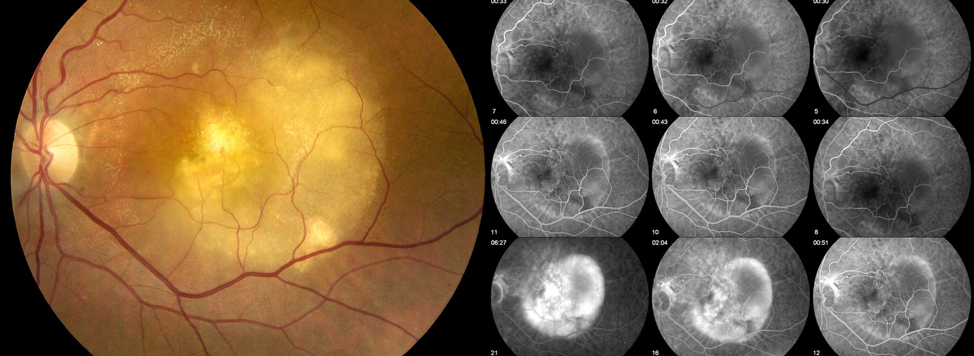
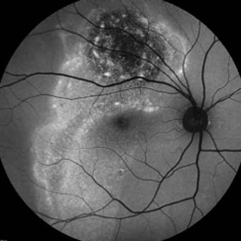
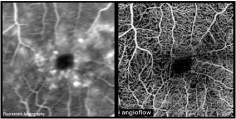
Fluorescentie Angiografie
Fluorescentie angiografie is een onderzoeksmethode waarbij foto's van het netvlies worden gemaakt met behulp van blauw flitslicht en een speciaal fototoestel.
Een in water oplosbare kleurstof (fluoresceïne of infracyanine groen (ICG)) wordt in een ader in de arm gespoten.
De kleurstof verspreidt zich via de grote lichaamsader door het hele lichaam en wordt dus ook naar het oog getransporteerd. De kleurstof verspreidt zich vrij snel na inspuiting door de bloedvaten van het oogvlies. Er worden dan meerdere foto's van het netvlies gemaakt.
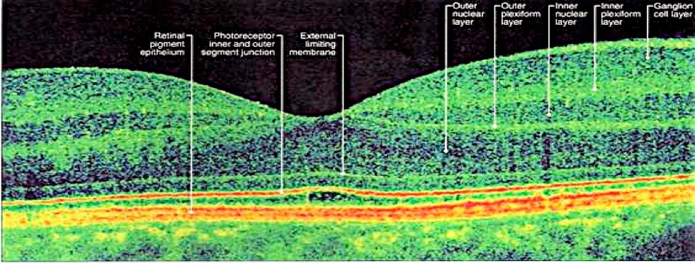
Optical Coherance Tomography - OCT
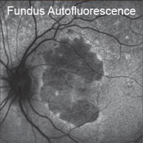
Autofluorescentie onderzoek - FAF
Bij het FAF onderzoek worden van het oplichtende netvlies NA het flitslicht.
Deze autofluorescentie wordt veroorzaakt door het lipofuscine in het retina pigment epitheel (RPE). Dit RPE blad is de buitenste laag van het netvlies. Lipofuscine geeft spontaan licht af na te zijn belicht met een felle lichtbron zoals een electronenflits.
Dit onderzoek geeft informatie over het metabolisme (de stofwisseling) en de functie van het retina pigment epitheel.
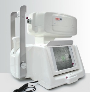
The MAIA software, with its normative database and statistical analysis module, is designed to identify the normal, age‐related, decrease in sensitivity and differentiate it from the pathological changes associated with macular degenerations and other retinal diseases.
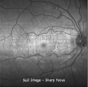
By using a line scanning laser ophthalmoscope (LSLO) to continuously image the retina, the light reflected by the out‐of‐focus layers does not degrade the quality of the image. The sharp constrast of the SLO image is due to its confocal properties.
EYE TRACKER
The role of the eye tracker is to compensate for the patient’s ocular movements during testing to ensure that point‐to‐point correspondence exists between the stimulus and the measured retinal location during the test and on subsequent tests.
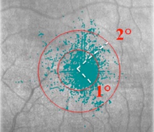
The automated eye tracker is fast, accurate and robust. It locks onto the entire fundus image and captures fixation changes 25 times per second during testing. Live images of the retina are processed to “track” anatomical landmarks over the entire image and quantify the corresponding eye movements observed during the test procedure. The software also identifies the Preferred Retinal Locus (PRL) – a retinal position that identifies the patient’s preferred fixation site. This is the location around which the perimetric stimuli will be projected.
MICROPERIMETRY
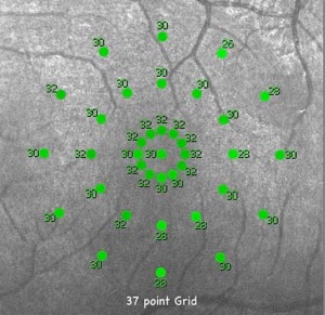
Measured values areaveraged and compared with the age‐adjusted average for normals. The average macular threshold, along with other variables, serves for the calculation of the macular integrity index, a statistical parameter derived by comparison with normative data that describes the likelihood of threshold values to significantly differ from normal values.
De Lairessestraat 59 1071 NT Amsterdam 020-679 71 55 omca@me.com www.omca.nl
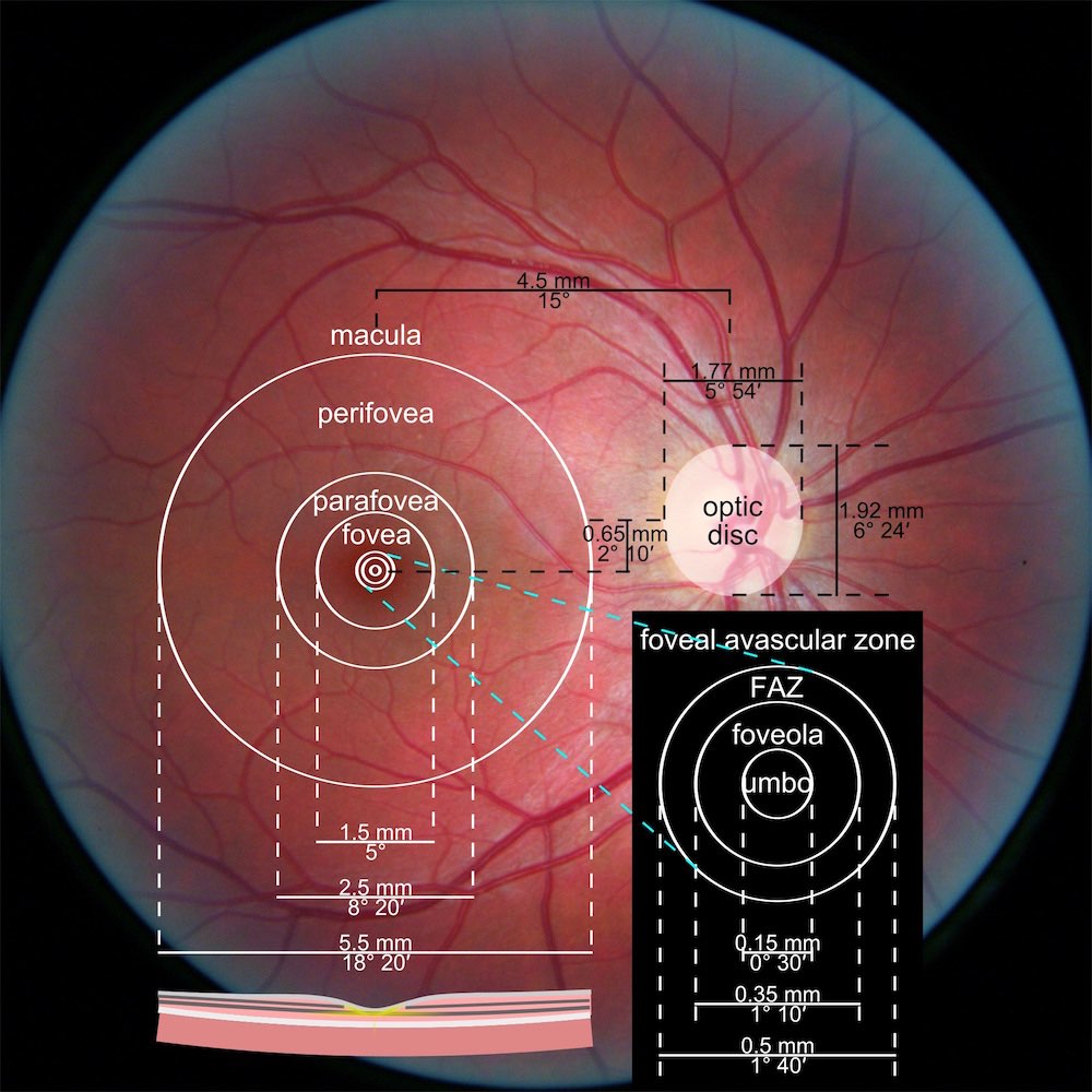
Macular Integrity Assesment - MAIA
De OptoVue Avanti is de nieuwe standaard voor macula onderzoek


Amsterdam Eye Hospital
Oogziekenhuis Amsterdam