MAIA offers three unique testing procedures – a fast supra‐threshold analysis, an expert or detailed threshold test and a follow‐up threshold analysis. These tests are designed to quantify macular threshold sensitivity and fixation stability and once an abnormal result is identified, to monitor those parameters over time.

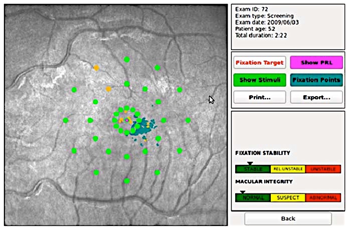
The Fast test is a supra‐threshold assessment to quickly evaluate macular sensitivity.
By performing 1‐2 projections at each pre‐defined retinal location and by processing the point‐ results to derive a combined evaluation of all measured points, macular integrity is categorized as normal, suspect or abnormal.
Fixation stability is judged as stable, relatively unstable or unstable based on the data obtained from the eye tracking. The preferred retinal locus (PRL) and fixation points can be displayed as needed.
The entire Fast test procedure takes around 2 minutes per eye. Patients with either suspect or abnormal results on the Fast test should have the Expert test
performed to accurately quantify retinal threshold and fixation stability. Other tests, including dilated fundus examination, optical coherence tomography, conventional fundus imaging and visual acuity testing, may be required to understand the cause of the abnormal findings.
The Fast test may be performed annually for those patients with a family history of a first‐degree relative with macular degeneration, unexplained vision loss, diabetes, post operative cataract surgery and cystoid macular edema.
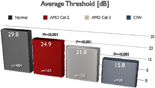
The sensitivity of the Fast test to reveal the alterations typical of Age related Macular Degeneration (AMD) hasbeen clinically assessed to be 90.7% and its specificity 90.1% (N = 813).

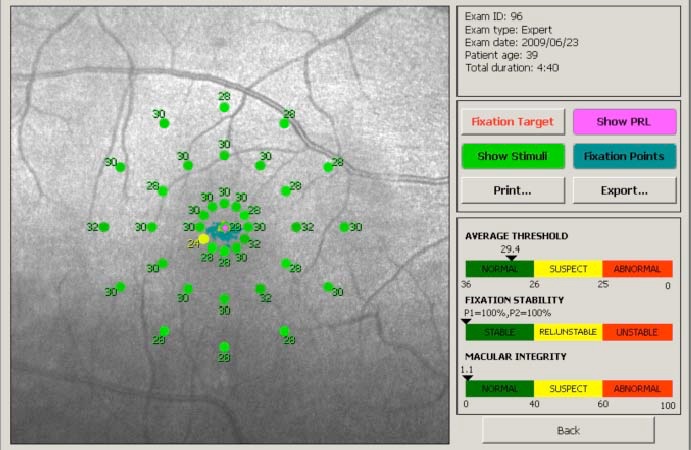
The Expert test should be performed to precisely quantify the extent and severity of functional visual loss associated with retinal disease. It is used to measure progressive changes in macular threshold sensitivity and fixation stability.
The Expert test uses a 4‐2 staircase to measuremacular threshold at each of the pre‐determinedpoints.
As a full threshold test, it takes around 5 minutes per eye to complete. Test outputs include: threshold values in dB at all measured points, average threshold sensitivity in dB, color coded according to the normality ranges, fixation stability index (the percentage of fixation points within 2° and 4° from the center of fixation) and macular integrity index, indicating the likelihood of presence of a statistically significant alteration in macular sensitivity when compared with normal values.
The preferred retinal locus (PRL) and fixation points can be displayed as needed. The sensitivity of the Expert test to reveal the alterations typical of AMD has been clinically assessed to be 91.2% and its specificity 98.4% (N = 813).

The follow‐up test is identical to the expert test, in that measured points are located at the same anatomical areas as in the baseline Expert test. The follow‐up test starts the threshold intensity for each point at the last recorded threshold obtained from the previous Expert test. This is designed to shorten the required time to take the test.
De Lairessestraat 59 1071 NT Amsterdam 020-679 71 55 omca@me.com www.omca.nl


Amsterdam Eye Hospital
Oogziekenhuis Amsterdam

