

Optic Nerve Glioma Resection
De Lairessestraat 59 1071 NT Amsterdam 020-679 71 55 omca@me.com www.omca.nl
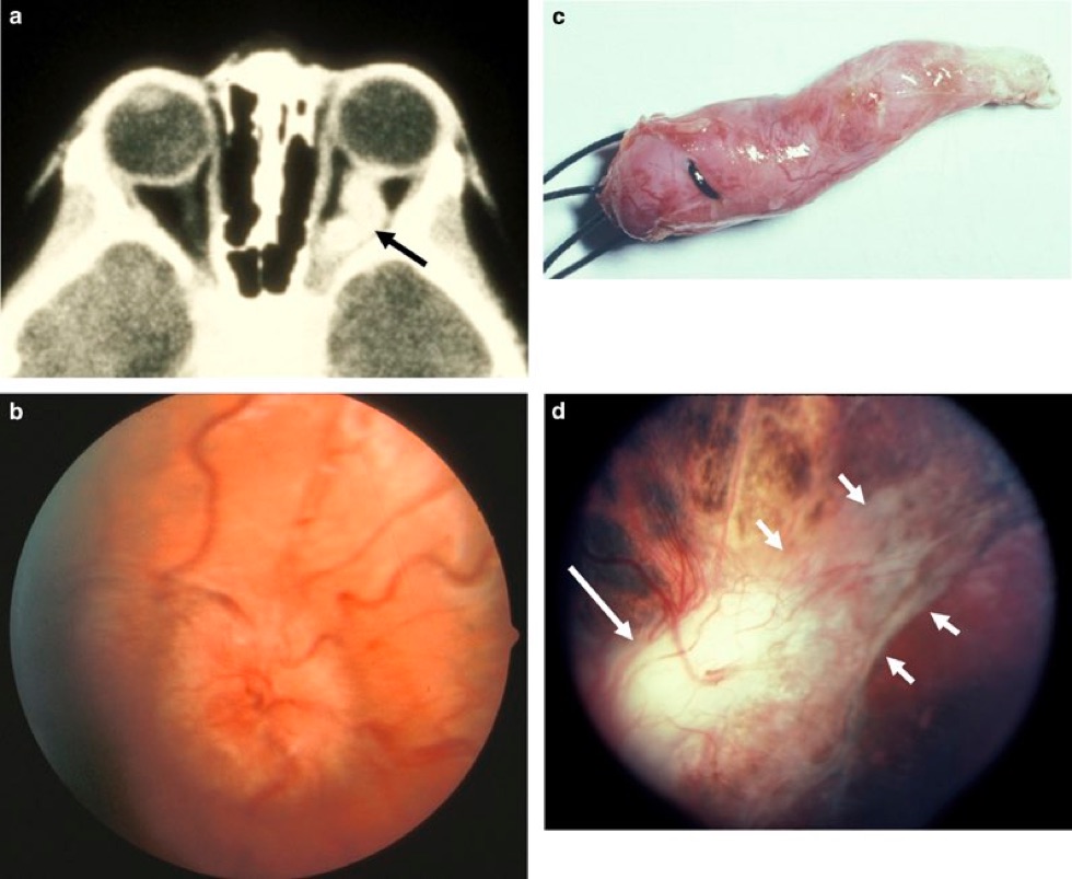
(a) Axial orbit CT scan with contrast of patient #4 obtained at the age of 39 months shows enhancing glioma involving the left intraorbital optic nerve (arrow). (b) Fundus photograph of patient #4 at the age of 39 months. Image shows marked swelling of the left optic disc. (c) Patient #4 was treated with an en-bloc excision of the orbital optic glioma of the left eye. Photograph shows the excised specimen. (d) Fundus photograph of the left eye of patient #4, 3 months after the optic nerve excision. Image shows optic disc pallor (long arrow) and tractional retinal detachment (short arrows).


Amsterdam Eye Hospital
Oogziekenhuis Amsterdam