

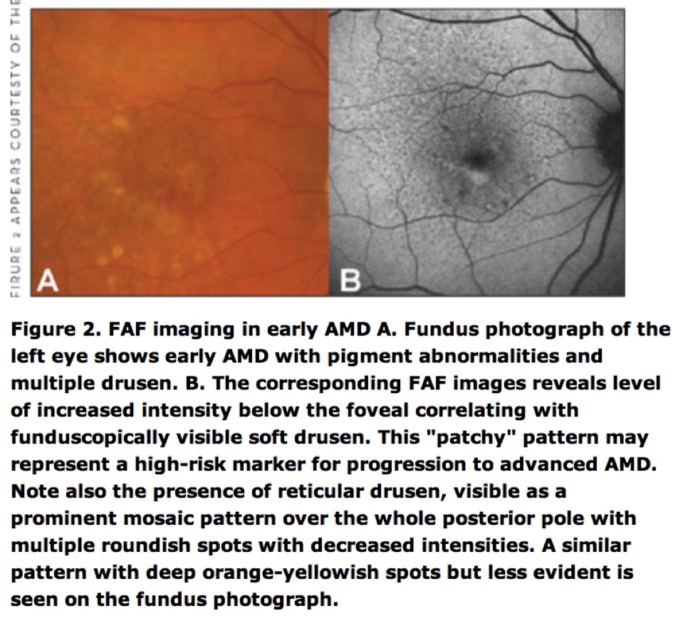
FAF AMD #1
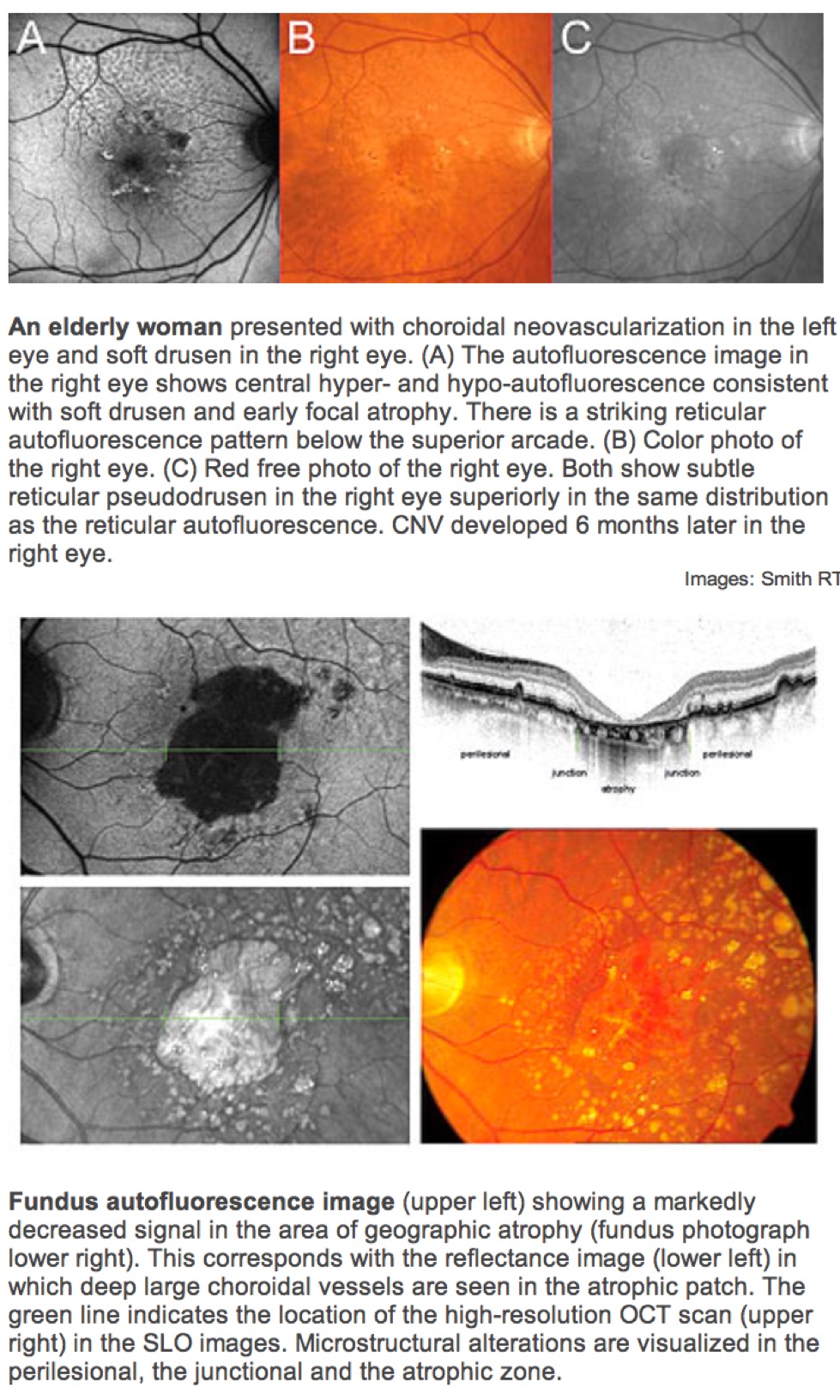
Illustration of advanced atrophy age-related macular degeneration in the left eye of a 55-year-old man.
A: Fundus photograph.
B: Fundus autofluorescence (FAF) image.
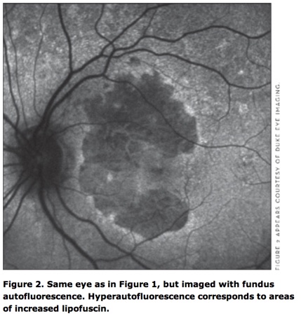
In the FAF image, the intensity of the large, kidney-shaped atrophic area in the center is markedly decreased because of the loss of lipofuscin accumulation and can be clearly delineated on the FAF image.
The surrounding retinal area looks normal on the fundus photograph, whereas the FAF images gives additional information showing increased FAF intensities around atrophy.
The fundus autofluorescence images at the bottom (C) demonstrate atrophy progression over time (6 years from baseline). Existing atrophy enlarges or new atrophy occurs exactly there where previously increased FAF had been observed.
De Lairessestraat 59 1071 NT Amsterdam 020-679 71 55 omca@me.com www.omca.nl


Amsterdam Eye Hospital
Oogziekenhuis Amsterdam