Effect op de bloedvaten


EFFECTS OF GLAUCOMA ON OCULAR STRUCTURES
Vasculature
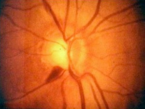
Several retinal vessel signs may suggest glaucoma. Vessel kinking/bayoneting at the cup edge is a classic finding due to the undermining of the neuroretinal rim tissue in glaucoma.
Nasal migration of disc vessels may occur over time, although this may or may not be a reliable indicator of glaucoma.
Vessel “bowing” in cup may help determine cup depth and size.
Baring of circumlinear vessels occurs as glaucoma advances due to exposure of the retinal arterioles and venules that reside in the gradually atrophying nerve fiber layer.
As the nerve fibers die, the vessels become more exposed creating sharper appearing vessels.
Other vascular changes that occur in glaucoma include overpass cupping in which vessels eventually collapse into a bared area of atrophy in the optic cup.
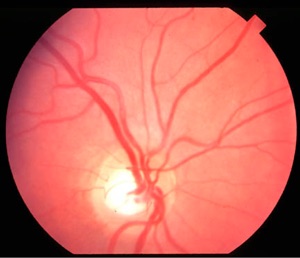
Drance splinter hemorrhages (flame-shaped hemorrhage crossing the optic nerve edge, usually at the inferior temporal disc margin). Because Drance hemorrhages precede glaucomatous damage in the nerve fibers, they are an ominous prognostic sign.
Drance (splinter) hemorrhage at 7:00 o'clock associated with normal-tension glaucoma.
Spontaneous venous pulsation can be present in normals, but arteriolar pulsation usually indicates excessively high intraocular pressure, or excessively reduced vascular pressure.
De Lairessestraat 59 1071 NT Amsterdam 020-679 71 55 omca@me.com www.omca.nl


Amsterdam Eye Hospital
Oogziekenhuis Amsterdam