Effect op de zenuwvezellaag


De Lairessestraat 59 1071 NT Amsterdam 020-679 71 55 omca@me.com www.omca.nl
EFFECTS OF GLAUCOMA ON OCULAR STRUCTURES
Nerve Fiber Layer
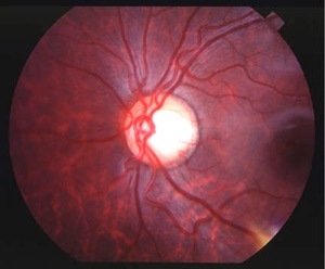
Superior and inferior nerve fiber layer striations in the retina of a Black patient.
Examination of the nerve fiber layer can be performed with a clear binocular indirect or high plus lens. However, a red-free filter can highlight its appearance.
The nerve fiber layer is the thickest in the inferior and superior areas, due to the neurons arching around the macula coming from the temporal periphery (papilomacular bundle) in addition to the neurons coming from the inferior and superior periphery.
Defects in the nerve fiber layer may be seen as dark slits or wedges that emanate from optic nerve margin.
An inferior temporal wedge defect is the most obvious nerve fiber layer defect and usually corresponds with inferior temporal notching and superior visual field loss. Slit defects (slightly larger than arterioles at the disc) may also be present although these can be seen in about 10% of normals.
Diffuse nerve fiber layer thinning is more common in high pressure glaucoma and corresponds to diffuse visual field loss and constriction.
Of interest, “rake” nerve fiber layer defects associated with small refractile talc bodies may indicate that the patient has a history of injected substance abuse (e.g., cocaine or heroin). Early glaucomatous-like paracentral scotomas may also appear in the visual field of these individuals, confusing the presentation with glaucoma.


Amsterdam Eye Hospital
Oogziekenhuis Amsterdam